Home
/ Blood Vessels Labeled : Blood Vessel Man Model Youtube - Arm blood vessels labeled :
Blood Vessels Labeled : Blood Vessel Man Model Youtube - Arm blood vessels labeled :
Blood Vessels Labeled : Blood Vessel Man Model Youtube - Arm blood vessels labeled :. Based on their structure and function, blood vessels are classified as either arteries, capillaries, or veins. The inner lining is the endothelium and is. Figures 1 and 2 show the major arteries and veins of the body. The word vascular, meaning relating to the blood vessels, is derived from the latin vas, meaning vessel. Deep veins, located in the center of the leg near the leg bones, are enclosed by muscle.
The blood vessels labeled h are called glomerulus and interact with the nephron to remove aged blood cells from the circulatory system * this problem has been solved! The vessels that carry blood away from the heart are called arteries, and their very small branches are arterioles. Blood vessels can be damaged by the effects of high blood glucose levels and this can in. This article lists a series of labeled imaging anatomy cases by system and modality. Very small branches that collect the blood from the various organs and parts are called venules, and they unite to form veins, which return the blood to the heart.
Honors Anatomy Chapter 11 Unit 3 Blood Vessels And Circulation Diagram Quizlet from o.quizlet.com The systemic blood vessels present in the thorax include: A primary purpose and significant role of the vasculature is its participation in oxygenating the body. Human cadaver, anatomical models, histology, cat, and fetal pig. The vessels that carry blood away from the heart are called arteries, and their very small branches are arterioles. The three major types of blood vessels: Based on their structure and function, blood vessels are classified as either arteries, capillaries, or veins. Compare fetal circulation to that of an individual after birth; Structure & function of blood vessels.
Structure & function of blood vessels.
Cat blood vessels labeled / major arteries and veins of the cat | anatomy corner. Veins (in blue) are the blood vessels that return blood to the heart. The inner lining is the endothelium and is. The common cartoid artery extends from the brachiocephalic artery. Blood vessels can be damaged by the effects of high blood glucose levels and this can in. Deoxygenated blood from the peripheral veins is transported back to the heart from capillaries, to venules, to veins, to the right side of the heart, and then. As the abdomen and pelvis contain the majority of internal organs, these regions need to be supplied by an extensive network of arteries and veins. Figures 1 and 2 show the major arteries and veins of the body. Blood pressure is measured as two readings, systolic and diastolic. Blood vessels form a continuous path for blood flow that starts and ends at the heart.arteries carry blood away from the heart, regardless of the degree of blood oxygenation.veins carry blood toward the heart. Compare fetal circulation to that of an individual after birth; Arm blood vessels labeled : Blood vessel labeling 9p image quiz.
Blood vessel labeling 7p image quiz. Between arteries and veins, there is a network of. This set is often in folders with. Eventually, the smallest arteries, vessels called arterioles, further branch into tiny capillaries, where nutrients and wastes are exchanged, and then combine with other vessels that exit capillaries to form venules, small blood vessels that carry blood to a vein, a larger blood vessel that returns blood to the heart. Identify and describe the hepatic portal system;
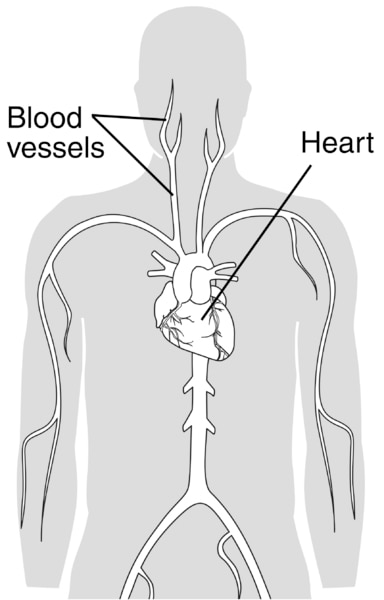
Torso With The Heart And Blood Vessels Labeled Media Asset Niddk from www.niddk.nih.gov The walls of blood vessels differ depending on the type of vessel. That being said, all arterial blood delivered to this region comes via branches of the abdominal aorta, and all venous blood eventually finds its way back to. A primary purpose and significant role of the vasculature is its participation in oxygenating the body. Arm blood vessels labeled : Describe the development of blood vessels and fetal circulation; •formed where capillaries unite • extremely porous 1) venules: Blood vessel structure and function. All blood vessels are basically hollow tubes with an internal space, called a lumen, through which blood flows.
Deoxygenated blood from the peripheral veins is transported back to the heart from capillaries, to venules, to veins, to the right side of the heart, and then.
These are discussed individually, below. Veins return blood back toward the heart. Blood vessels of the abdomen and pelvis. That being said, all arterial blood delivered to this region comes via branches of the abdominal aorta, and all venous blood eventually finds its way back to. Spend a while piecing these. The word vascular, meaning relating to the blood vessels, is derived from the latin vas, meaning vessel. The vessels that carry blood away from the heart are called arteries, and their very small branches are arterioles. Describe the development of blood vessels and fetal circulation; Blood vessels 2 labeled palmar arch digital artery right femoral a right femoral v great saphenous vein left popliteal a right anterior tibial a. This article lists a series of labeled imaging anatomy cases by system and modality. Veins (in blue) are the blood vessels that return blood to the heart. What is the name of blood vessel b? Eventually, the smallest arteries, vessels called arterioles, further branch into tiny capillaries, where nutrients and wastes are exchanged, and then combine with other vessels that exit capillaries to form venules, small blood vessels that carry blood to a vein, a larger blood vessel that returns blood to the heart.
Vessels transport nutrients to organs/tissues and to transport wastes away from organs/tissues in the blood. Arteries, veins, and capillaries blood vessels flow blood throughout the body. This set is often in folders with. Blood vessel labeling 7p image quiz. That being said, all arterial blood delivered to this region comes via branches of the abdominal aorta, and all venous blood eventually finds its way back to.
Wire Models from classroom.sdmesa.edu Human cadaver, anatomical models, histology, cat, and fetal pig. The major arteries in the body. Blood circulates throughout the body in blood vessels, propelled by the pumping action of the heart. The adventitia or outer layer which provides structural support and shape to the vessel •formed where capillaries unite • extremely porous 1) venules: The word vascular, meaning relating to the blood vessels, is derived from the latin vas, meaning vessel. Professor fink reviews cat blood vessels; Blood vessel labeling 7p image quiz.
Aorta, brachiocephalic trunk, brachiocephalic veins, superior and inferior vena cava, azygous vein and the vertebral veins.
Hma practical 3 for monday july 23 and wednesday july 25. As the abdomen and pelvis contain the majority of internal organs, these regions need to be supplied by an extensive network of arteries and veins. Dr calum worsley and assoc prof craig hacking et al. Blood vessels form a continuous path for blood flow that starts and ends at the heart.arteries carry blood away from the heart, regardless of the degree of blood oxygenation.veins carry blood toward the heart. The inner lining is the endothelium and is. Blood vessels of the abdomen and pelvis. These are discussed individually, below. Veins return blood back toward the heart. Blood vessel labeling 7p image quiz. Identify and describe the hepatic portal system; Deep veins, located in the center of the leg near the leg bones, are enclosed by muscle. The common cartoid artery extends from the brachiocephalic artery. Deep veins, located in the center of the leg near the leg bones, are enclosed by muscle.

